Home
/ Plant Cell Microscope Organelles : Microscopy Intro To Microscopes How They Work Article Khan Academy / In plant cells, the cell membrane lies inside the cell wall.
Plant Cell Microscope Organelles : Microscopy Intro To Microscopes How They Work Article Khan Academy / In plant cells, the cell membrane lies inside the cell wall.
Plant Cell Microscope Organelles : Microscopy Intro To Microscopes How They Work Article Khan Academy / In plant cells, the cell membrane lies inside the cell wall.. For only $16.38 $13.9 /page. They coordinate and function efficiently for the normal functioning of the cell. Animal and plant cells undergo a precise type of division called mitosis. By looking at the slide of rice leaf, you can see the vascular system extending from the stem into the leaves as a continuous pipe network. What organelles could not be seen?
They must draw and label the nucleus, cell membrane, cell wall, vacuole, chloroplasts. The figure below is a fine structure of a generalized animal cell. What is the composition of a plant cell wall? Closeup views and animations of certain organelles is provided. Students will observe cheek cells under a microscope.
Organelles Join Mitochondria In Signalling Pathways Creation from scx2.b-cdn.net It must have been a light microscope or tem. Cell wall and cell membrane a. Ability to explain how the composition of organelles will be different in cells with different functions, (e.g. This appears at the light microscope level as a duplication of chromosomes. The organelles that are unique for plant cells are vacuole, cell wall, and chloroplast (shown in orange text). They must draw and label the nucleus, cell membrane, cell wall, vacuole, chloroplasts. Discuss the tools and methods for viewing cells. Cell staining is a technique that can be used to visualize cells and cell components under a microscope.
Students will observe onion cells under a microscope.
They must draw and label the nucleus, cell membrane, cell wall, vacuole, chloroplasts. Discuss the tools and methods for viewing cells. Plant cells are eukaryotic cells whose cells have membrane bound nuclei and organelles. They coordinate and function efficiently for the normal functioning of the cell. Elodea are common freshwater aquarium plants. The photograph shown below details chloroplast structure as viewed with a transmission electron microscope courtesy of dr. Fill out the table below to show what we could and could not see through our microscope. Explain how the structure of prokaryotes allows them to divide by binary fission. Photosynthetic cells of the leaf of elodea. Generalized cell is used for structure of animal cell and plant cell to present the common parts, appearing in various parts of the bodies of animals and plants. Start studying 6.1 microscope and cell organelles. We will write a custom essay sample on microscope observation of animal and plant cells specifically for you. What organelles could not be seen?
What organelles could be seen? The function of the leaves is to collect energy from the sunlight and convert the energy into sugars for the plant. What organelles could be seen? What is the composition of a plant cell wall? Animal and plant cells undergo a precise type of division called mitosis.
Confocal Laser Scanning Microscopy Of Plant Cells Labeled With Various Download Scientific Diagram from www.researchgate.net The cell wall of a plant is made up of cellulose. Animal and plant cells undergo a precise type of division called mitosis. Students will observe onion cells under a microscope. The organelles that are unique for plant cells are vacuole, cell wall, and chloroplast (shown in orange text). If the cell is very tiny, then a tem was used. Fill out the table below to show what we could and could not see through our microscope. It must have been a light microscope or tem. When seen under a microscope, a plant cell is somewhat rectangular in shape and displays a double membrane which is more rigid than that of an animal cell.
Plant and animal cells microscope lab.
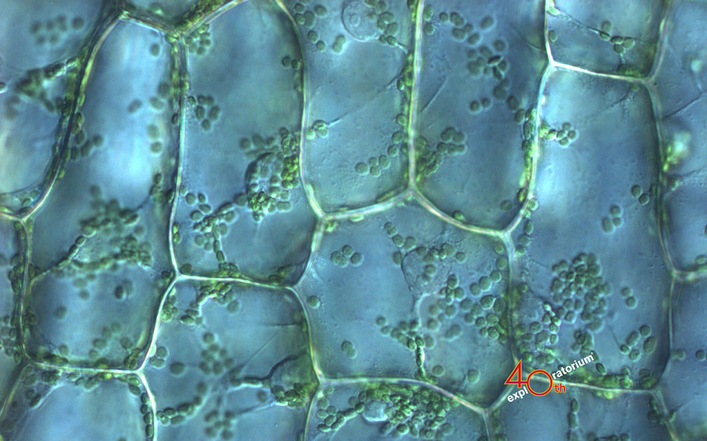
Chromatin nucleus chromatin the vacuole in this mature plant cell from a leaf is large, and occupies about 80% of the cell volume. The plant cell back to menu or next or previous. Students will observe cheek cells under a microscope. The cell wall, large central vacuole and chloroplasts are clearly visible. Before cell division, the entire genome is copied. When students finish their diagrams and have their drawing approved, they move on to an assigned microscope. Photosynthetic cells of the leaf of elodea. Plant and animal cells microscope lab. Endoplasmic reticulum rough and smooth british society for. Ability to explain how the composition of organelles will be different in cells with different functions, (e.g. An elodea leaf was mounted in pondwater between a slide and coverslip with a silicon spacer. Start studying 6.1 microscope and cell organelles. For the plant section, students should note that plant cells have chloroplasts, cell walls, and larger vacuoles.
The chloroplasts contain a green pigment chlorophyll that is responsible for the process of photosynthesis. It must have been a light microscope or tem. Photosynthetic cells of the leaf of elodea. Students will discover that their skin is made up of cells. Discuss the tools and methods for viewing cells.
Plant Cell Structure Plant Cell Parts Organelles And Their Functions And Diagram Jotscroll from www.jotscroll.com The chloroplasts contain a green pigment chlorophyll that is responsible for the process of photosynthesis. The figure below is a fine structure of a generalized animal cell. In plant cells, the cell membrane lies inside the cell wall. Images were taken on an inverted compound microscope using a 40x dic objective and digital camera. What organelles could be seen? When students finish their diagrams and have their drawing approved, they move on to an assigned microscope. Elodea are common freshwater aquarium plants. Animal and plant cells have certain.
What are the differences between a plant and animal cell?
What is the composition of a plant cell wall? By looking at the slide of rice leaf, you can see the vascular system extending from the stem into the leaves as a continuous pipe network. Three layers together measure approximately 75 å in thickness. Cell wall and cell membrane a. Fill out the table below to show what we could and could not see through our microscope. After, they will engage in a reading assignment, worksheets, and discussion to discover how the two types of cells are similar and different and what the role of the organelles are. In most plant cells, the organelles that are visible under a compound light microscope are the cell wall, cell membrane, cytoplasm, central vacuole, and nucleus. Students will discover that their skin is made up of cells. The animal cell and plant cell share many organelles in common, such as a nucleus, er, cytosol, lysosomes, golgi apparatus, cell membrane, and ribosomes. The plant cell back to menu or next or previous. Animal and plant cells have certain. It must have been a light microscope or tem. Plant and animal cells microscope lab.
Share :
Post a Comment
for "Plant Cell Microscope Organelles : Microscopy Intro To Microscopes How They Work Article Khan Academy / In plant cells, the cell membrane lies inside the cell wall."


Post a Comment for "Plant Cell Microscope Organelles : Microscopy Intro To Microscopes How They Work Article Khan Academy / In plant cells, the cell membrane lies inside the cell wall."