Home
/ Diagram Of Plant Cell As Seen Under Light Microscope : Components Teacher Stem In Curriculum Mr Mwaurah School / Plant cells are the basic unit and building blocks of life in organisms of the kingdom plantae.
Diagram Of Plant Cell As Seen Under Light Microscope : Components Teacher Stem In Curriculum Mr Mwaurah School / Plant cells are the basic unit and building blocks of life in organisms of the kingdom plantae.
Diagram Of Plant Cell As Seen Under Light Microscope : Components Teacher Stem In Curriculum Mr Mwaurah School / Plant cells are the basic unit and building blocks of life in organisms of the kingdom plantae.. Cells consist of cytoplasm enclosed within a membrane, which contains many biomolecules such as proteins and nucleic acids.2 most plant and animal cells are only visible under a light microscope, with dimensions between 1. The term 'cell' was coined to describe the small walled units that were observed in the sections of bottle cork under simple microscope. Here's a photo of a plant cell under an electron microscope. To use a light microscope to examine animal or plant cells. Chloroplasts are organelles found in the cytoplasm that are packed endoplasmic reticulum studded with ribosomes looks rough under the microscope;
The study of cells is called cell biology, cellular biology, or cytology. Describe and compare the structure of a plant cell with an animal cell, as seen under a light microscope, limited to cell wall, nucleus, cytoplasm, chloroplasts, vacuoles and location of the cell membrane. One can visualize molecules and even atoms using an electron. Inulin is spherical or star shaped crystal. The cell wall is made of cellulose and surrounds the cell membrane in plant cells.
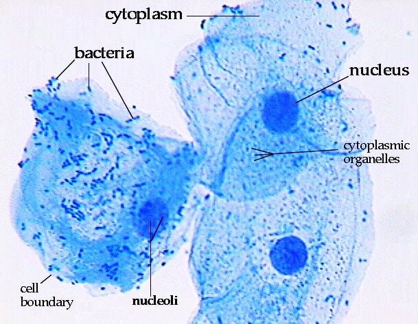
Cape Biology And Chemistry Syllabus Biology Module 1 2 Cell Structure 2 1 Make Drawings Of Typical Animal And Plant Cells As Seen Under The Light Microscope from download.e-bookshelf.de The cell wall is made of cellulose and surrounds the cell membrane in plant cells. The plant cell as more rigid and stiff walls. Cells of plant or animal tissue. Features of cells are possessed by all living things? The plant cell is the basic structural and functional unit found in the members of the kingdom plantae. They are cells that have a distinct nucleus and other cellular organelles under the microscope, it shows many different parts. Under a light microscope, the cell membrane, nucleus and cytoplasm of a cheek cell (animal cell) can be observed. A scale bar has been marked on the drawing, allowing the.
The cell wall is made of cellulose and surrounds the cell membrane in plant cells.
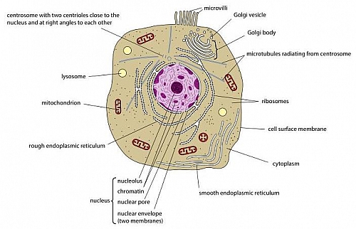
They are green in color under a microscope because they. Chloroplasts are organelles found in the cytoplasm that are packed endoplasmic reticulum studded with ribosomes looks rough under the microscope; The term 'cell' was coined to describe the small walled units that were observed in the sections of bottle cork under simple microscope. Diagram 1 shows the structure of a plant cell as seen under light microscope. Given below is the diagram of a cell as seen under the microscope after having been placed in a solution What was once unseeable can now be seen, touched, and eaten!cut. Rayliq is waiting for your help. This is the reason why you need to use a microscope to observe a cell. (iii) presence of cell wall. Magnification, however, is not the most important issue in microscopy. (ii) presence of large central vacuole in plant cell. Cells are generally microscopic, so you need a microscope to see the parts they are made of. Plant cells have cell walls, one large vacuole per cell, and chloroplasts, while animal cells will have a cell membrane only.
Diagram 1 shows the structure of a plant cell as seen under light microscope. Cells are generally microscopic, so you need a microscope to see the parts they are made of. This is a single cheek cell that has been stained, as seen under a light microscope. Chloroplasts are organelles found in the cytoplasm that are packed endoplasmic reticulum studded with ribosomes looks rough under the microscope; The plant cell as more rigid and stiff walls.
Animal Cell Structure And Organelles With Their Functions Jotscroll from www.jotscroll.com The plant cell is the basic structural and functional unit found in the members of the kingdom plantae. Magnification, however, is not the most important issue in microscopy. Plant cells have cell walls, one large vacuole per cell, and chloroplasts, while animal cells will have a cell membrane only. Features of cells are possessed by all living things? This site is using cookies under cookie policy. They are green in color under a microscope because they. Animal cells also have a because only plant cells perform photosynthesis, chloroplasts are found only in plant cells. Plant cells do, however, have a number of other specialized structures, including a rigid cell wall, central vacuole, plasmodesmata, and chloroplasts.
Magnification, however, is not the most important issue in microscopy.
Plant cell under electron microscope. Here's a photo of a plant cell under an electron microscope. Absorption of water by the root hair of plant absorption of glucose through the villi in the small. To make observations and draw scale. Click (or tap) the diagram for a simple labelled version. Resolving power is the ability to distinguish between separate things which are close to each other. They are green in color under a microscope because they. To use a light microscope to examine animal or plant cells. Ishita observed a slide of eukaryotic cell under electron microscope. One can visualize molecules and even atoms using an electron. Light microscopes using visible light and lenses to form a magnified image of the object under investigation e.g. The diagram below is a plant cell as may be seen using a light microscope. Describe and compare the structure of a plant cell with an animal cell, as seen under a light microscope, limited to cell wall, nucleus, cytoplasm, chloroplasts, vacuoles and location of the cell membrane.
Plant cell science diagram clipart set includes: Draw a table of differences between the microscopes are instruments that are used to look at and study objects that are too small to be when we view a specimen under a microscope it needs to let light pass through the specimen so. Here's a diagram of a plant cell: A scale bar has been marked on the drawing, allowing the. It also has a very high resolving power.
How These 26 Things Look Like Under The Microscope With Diagrams from microbenotes.com This is the reason why you need to use a microscope to observe a cell. The compound light microscope operates on the main principle that an objective lens with a very short focal length can form a highly magnified real image of the object. The labels in pink are links to pages of further information. This gives rise to its name of rough endoplasmic reticulum. Some of these differences can be clearly understood when the cells are examined under an electron microscope. One can visualize molecules and even atoms using an electron. Plant cells have cell walls, one large vacuole per cell, and chloroplasts, while animal cells will have a cell membrane only. To make observations and draw scale.
You can specify conditions of storing and accessing cookies in your browser.
Chloroplasts are organelles found in the cytoplasm that are packed endoplasmic reticulum studded with ribosomes looks rough under the microscope; Plant cell under electron microscope. Cells are generally microscopic, so you need a microscope to see the parts they are made of. Under a light microscope, the cell membrane, nucleus and cytoplasm of a cheek cell (animal cell) can be observed. Describe and compare the structure of a plant cell with an animal cell, as seen under a light microscope, limited to cell wall, nucleus, cytoplasm, chloroplasts, vacuoles and location of the cell membrane. The cell wall is made of cellulose and surrounds the cell membrane in plant cells. Rayliq is waiting for your help. Vpc 360° video by plant energy biology. Ishita observed a slide of eukaryotic cell under electron microscope. This gives rise to its name of rough endoplasmic reticulum. This site is using cookies under cookie policy. Learn about the size and function of plant and animal cells for gcse combined science once slides have been prepared, they can be examined under a microscope. Here's a diagram of a plant cell:
Share :
Post a Comment
for "Diagram Of Plant Cell As Seen Under Light Microscope : Components Teacher Stem In Curriculum Mr Mwaurah School / Plant cells are the basic unit and building blocks of life in organisms of the kingdom plantae."


Post a Comment for "Diagram Of Plant Cell As Seen Under Light Microscope : Components Teacher Stem In Curriculum Mr Mwaurah School / Plant cells are the basic unit and building blocks of life in organisms of the kingdom plantae."