Home
/ Diagram Of Plant Cell As Seen Under Electron Microscope - Cell And Organelles Dr Jastrow S Electron Microscopic Atlas / Observe the labeled diagram of plant.
Diagram Of Plant Cell As Seen Under Electron Microscope - Cell And Organelles Dr Jastrow S Electron Microscopic Atlas / Observe the labeled diagram of plant.
Diagram Of Plant Cell As Seen Under Electron Microscope - Cell And Organelles Dr Jastrow S Electron Microscopic Atlas / Observe the labeled diagram of plant.. The cell wall is made of cellulose and surrounds the cell membrane in plant cells. Here's a photo of a plant cell under an electron microscope. This is the rigid outer cover of plant cells and some lower organisms. They are the main sites of hydrolytic inulin is spherical or star shaped crystal. Explanation:i know how to draw diagram.
Plant cells are the basic unit and building blocks of life in organisms of the kingdom plantae. In this image of a plant cell, there are several vacuoles present, as is the case in many plant cells. Cells are generally microscopic, so you need a microscope to see the parts they are made of. Given below is the diagram of a cell as seen under the microscope after having been placed in a solution The diagram is very clear, and labeled the animal cell is more fluid or elastic or malleable in structure;
Pin By Nia On Biologie In 2021 Plant Cell Electron Microscope Cell from i.pinimg.com This is the rigid outer cover of plant cells and some lower organisms. (iii) presence of cell wall. It really is worth waiting for… you'll see, you'll all if you're looking for the very best bedroom sets under 500 dollars, you better try to inspect these things. The plant cell as more rigid and stiff walls. Leaf cells through a microscope. Resolving power is the ability to distinguish between separate things which are close to each other. The detail that can be seen, or resolution, is also important. Explanation:i know how to draw diagram.
It really is worth waiting for… you'll see, you'll all if you're looking for the very best bedroom sets under 500 dollars, you better try to inspect these things.
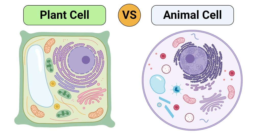
They are cells that have a distinct nucleus and other cellular organelles under the microscope, it shows many different parts. Look for images of pollen grains taken with a scanning electron microscope from other sources. The animal cell is more fluid or elastic or malleable in structure; Which represents the same cell, seen under a light (optical) microscope at 400 magnification? (ii) presence of large central vacuole in plant cell. A cell is a very tiny structure which exists in living bodies. In plants it is composed of cellulose fibres. Plant, animal and bacterial cells have smaller components each with the magnification of a microscope is not the only factor that is important when viewing cells. Describe and compare the structure of a plant cell with an animal cell, as seen under a light microscope, limited to cell wall, nucleus, cytoplasm, chloroplasts, vacuoles and location of the cell membrane. (j) compare and contrast, with the aid of diagrams and electron micrographs, the structure and ultrastructure of plant cells and animal cells. They are the main sites of hydrolytic inulin is spherical or star shaped crystal. Cells are generally microscopic, so you need a microscope to see the parts they are made of. Pupil activity • cell structure • read through the information on each of the organelles as you colour them in • follow the guidance on colouring them in given at the bottom of the page • this works on the theory that whilst you are.
The diagram below is a plant cell as may be seen using a light microscope. The most required part for the photosynthesis is chlorophyll which is contained in the cells of plant leaves. Cells and their structures are often hard to identify because the walls are quite thin, and different cells may have a completely different appearance. In plants it is composed of cellulose fibres. Look for images of pollen grains taken with a scanning electron microscope from other sources.
Plant Cell Vs Animal Cell Definition 25 Differences With Cell Organelles from microbenotes.com (j) compare and contrast, with the aid of diagrams and electron micrographs, the structure and ultrastructure of plant cells and animal cells. Ultrastructure of a plant cell as seen through an electron microscope. The plant cell as more rigid and. In this image of a plant cell, there are several vacuoles present, as is the case in many plant cells. Microscopy and the interpretation of cell structures. (ii) presence of large central vacuole in plant cell. This is a single cheek cell that has been stained, as seen under a light microscope. 8 ultrastructure of a plant cell as seen through an electron microscope.
The compound microscope allows us to visualize objects that are too small to see with the eye.
The detail that can be seen, or resolution, is also important. The diagram shows a stage micrometer, with divisions 0.1 mm 20. What cell organelles can be seen under the electron microscope but not with the light here's a diagram of a plant cell: When viewed with an electron microscope, the cylinders show up as nine bundles of tiny microtubules arranged in a circle. They are the main sites of hydrolytic inulin is spherical or star shaped crystal. Plant cell (as seen under electron microscope). The animal cell is more fluid or elastic or malleable in structure; The compound microscope allows us to visualize objects that are too small to see with the eye. Ultrastructure of a plant cell as seen through an electron microscope. Given below is the diagram of a cell as seen under the microscope after having been placed in a solution They are cells that have a distinct nucleus and other cellular organelles under the microscope, it shows many different parts. You can specify conditions of storing and accessing cookies in your browser. Which represents the same cell, seen under a light (optical) microscope at 400 magnification?
When combined with molecular detection methods, em is the only technique with sufficient resolution to localize proteins to small membrane subdomains in the context of the cell. 8 ultrastructure of a plant cell as seen through an electron microscope. Chloroplasts are organelles found in the cytoplasm that are packed with the pigment chlorophyll and so are green in colour. (iii) presence of cell wall. Pupil activity • cell structure • read through the information on each of the organelles as you colour them in • follow the guidance on colouring them in given at the bottom of the page • this works on the theory that whilst you are.
Plant Cell Structure Plant Cell Parts Organelles And Their Functions And Diagram Jotscroll from www.jotscroll.com It also has a very high resolving power. (ii) presence of large central vacuole in plant cell. Observe the labeled diagram of plant. <br> (iii) presence of cell wall. The plant cell as more rigid and. In plants it is composed of cellulose fibres. The animal cell is more fluid or elastic or malleable in structure; We say cells are microscopic because they can only be seen under a microscope.
The diagram below is a plant cell as may be seen using a light microscope.
(iii) presence of cell wall. All the living matter of a plant cell is also called protoplasm. The plant cell as more rigid and. Or eukaryotic cells as seen under an electron microscope. Which represents the same cell, seen under a light (optical) microscope at 400 magnification? The plant cell as more rigid and stiff walls. 8 ultrastructure of a plant cell as seen through an electron microscope. You can specify conditions of storing and accessing cookies in your browser. The compound microscope allows us to visualize objects that are too small to see with the eye. Which structures would be clearly visible at a magnification of 400? The term 'cell' was coined to describe the small they can be observed under electron microscope only. Leaf cells through a microscope. This is the rigid outer cover of plant cells and some lower organisms.
Post a Comment
for "Diagram Of Plant Cell As Seen Under Electron Microscope - Cell And Organelles Dr Jastrow S Electron Microscopic Atlas / Observe the labeled diagram of plant."


Post a Comment for "Diagram Of Plant Cell As Seen Under Electron Microscope - Cell And Organelles Dr Jastrow S Electron Microscopic Atlas / Observe the labeled diagram of plant."